Banner

Falar com
Dr. Marcelo

Sintomas iniciais e avançado do Ceratocone

Imagem Sintomas iniciais e avançado Ceratocone

A suspeita do ceratocone começa quando o paciente apresenta queixa de baixa visão e aumento progressivo do astigmatismo ou baixa visão mesmo com óculos. Nesses casos, muitas vezes o ceratocone já se encontra em um estágio evolutivo de moderado a avançado.

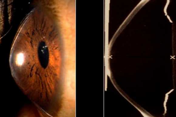
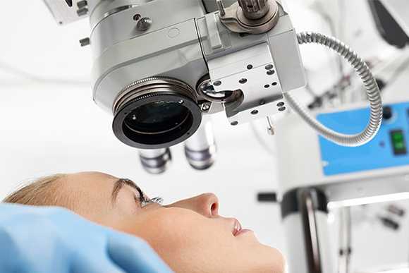
Atualmente, existem aparelhos mais específicos, capazes de detectar as formas iniciais do ceratocone (também chamados estágios subclínicos, quando o paciente ainda não apresenta sintomas importantes).

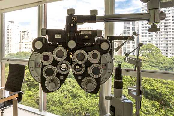
Esses aparelhos são capazes de medir com extrema precisão a curvatura e a espessura da córnea, bem como seu grau de irregularidade.

Imagem Visão sem e com Ceratocone
Visão Sem e Com Ceratocone

Com a contínua progressão do ceratocone, o astigmatismo aumenta bastante e se tornar irregular (gerando uma imagem borrada e distorcida), e os óculos passam a não mais oferecer uma visão satisfatória. Nesse estágio, somente as lentes de contato duras (rígidas) são capazes de melhorar parcialmente a visão.

Muitos pacientes apresentam uma estabilidade relativa ou lenta progressão do ceratocone nesse estágio e, se estiverem bem adaptados as lentes de contato rígidas, conseguem levar uma vida normalmente, sem muitas restrições.

Os pacientes não apresentam dor ou desconforto ocular nesses estágios iniciais da doença.

O aumento da curvatura (formato de cone) não pode ser visto a olho nu.

Em muitos casos, infelizmente, o ceratocone continua evoluindo progressivamente e a adaptação e tolerância as lentes de contato rígidas vão se tornando cada vez mais limitadas.

O aumento da protuberância na região central da córnea (cone) dificulta a estabilidade das lentes de contato, bem como diminui a visão do paciente. No estágio final do ceratocone, o transplante de córnea passa a ser a única alternativa terapêutica capaz de restabelecer parcialmente a visão.

O que é o Ceratocone

Imagem oque é Ceratocone

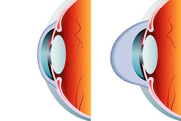
O ceratocone é uma doença da córnea, caracterizada pela aumento progressivo e irreversível de sua curvatura, bem como pela diminuição progressiva de sua espessura.

Em outras palavras, a córnea (parte mais anterior do olho), torna-se “pontuda” e “fina”.

Imagem Visão sem e com Ceratocone
Visão Sem e Com Ceratocone

Na evolução natural do ceratocone, a córnea perde progressivamente sua conformação original (levemente curva) e adquire um formato atípico (extremamente curva, semelhante a um cone).

O Que Causa o Ceratocone

Imagem O que causa Ceratocone

A causa exata do ceratocone ainda permanece desconhecida. Porém sabe-se que alguns genes estão diretamente envolvidos no aparecimento do ceratocone. Por isso, a presença do ceratocone é mais comum em membros de uma mesma família. Entretanto, sua evolução pode ser distinta entre os membros da família.

Sabe-se também que alguns fatores externos atuam acelerando a progressão do ceratocone, como traumas repetitivos durante o hábito de coçar os olhos cronicamente. Acredita-se também que a infecção por alguns vírus possa desencadear reações imunológicas capazes de estimular a progressão do ceratocone.

Quando aparece o Ceratocone?

Imagem Quando aparece o Ceratocone

O ceratocone começa a se desenvolver geralmente na adolescência, a partir dos 13 anos de idade e apresenta seu pico de evolução entre os 15 e 25 anos. Entretanto, existem alguns casos de aparecimento e evolução precoces (antes dos 15 anos) e também casos de aparecimento e evolução tardia (após os 30 anos).

Quem é o Dr. Marcelo. V. Netto

Imagem Dr. Marcelo V. Netto

Dr. Marcelo V. Netto – CRM (SP) 96405

O Dr. Marcelo é Especialista em Tratamento do Ceratocone.

Resumo Biografia:

  • Autor de 2 livros e 82 artigos científicos.
  • Prêmio Internacional Castroviejo Award da Sociedade Internacional de Cirurgia Refrativa
  • Prêmio Internacional Troutman Award da Academia Americana de Oftalmologia
  • O Dr. Marcelo tem Doutorado pela Universidade de São Paulo (USP).

Foi Senior Fellow e Instrutor Cirúrgico da Universidade de Washington, Seattle, EUA. Em seguida, concluiu o PhD em Ciências Visuais pela The Cleveland Clinic Foundation em Cleveland, OH, EUA.

Atualmente o Dr. Marcelo V. Netto está coordenando projetos de pesquisa no setor de córnea e cirurgia refrativa da Universidade de São Paulo (Hospital das Clínicas da Faculdade de Medicina da USP) em parceria com o IPEN (Instituto de Pesquisa e Energia Nuclear da USP) e The Cleveland Clinic Foundation, Ohio, US.

Sua produção científica inclui: Autor de 2 livros médicos na área de cirurgia refrativa e 82 artigos científicos publicados (veja abaixo relação dos principais artigos científicos publicados em revistas indexadas), mais de 50 capítulos de livros e autoria de dois livros “Atualização Propedêutica em Cirurgia Refrativa” e “Manual de Cirurgia Refrativa do Hospital das Clínicas da USP” .

PREMIAÇÕES

  • • Prêmio “Troutman Award” de Melhor artigo científico publicado no Journal of Refractive Surgery (Las Vegas, NV)
  • • Prêmio “Castroviejo Award” pela Cornea Society (New Orleans, LA)
  • • Prêmio “Achievement Award” pela American Academy of Ophthalmology (Atlanta, GA)
  • • Prêmio “Prêmio Founder’s Award” na IV Wavefront e Refractive Surgery Meeting (Nassau,Bahamas)
  • • Prêmio “Winner Video” pela American Society of Cataract and Refractive Surgery (Washington, D.C.)
  • • Prêmio “Best Poster” and “Best Paper” por dois anos consecutivos pela American Society of Cataract and Refractive Surgery (Washington, D.C. e San Diego, CA)
  • • Prêmio “Alumni Award”pela The Cleveland Clinic Foundation. (Cleveland, OH)

ARTIGOS PUBLICADOS

  • Very late-onset corneal scar triggered by trauma after photorefractive keratectomy. Gomes BA, Smadja D, Espana EM, Ahn ES, Netto MV, Santhiago MR. J Cataract Refract Surg. 2012 Sep;38(9):1694-7
  • Peripheral sterile corneal ring infiltrate after riboflavin-UVA collagen cross-linking in keratoconus. Ghanem RC, Netto MV, Ghanem VC, Santhiago MR, Wilson SE. Cornea. 2012 Jun;31(6):702-5.
  • Colírios Cicatrizantes: biological effects and use in refractive surgery. Santhiago MR, Netto MV, Wilson SE. Cornea. 2012 Mar;31(3):311-21.
  • Modulation transfer function and optical quality after bilateral implantation of a +3.00 D versus a +4.00 D multifocal intraocular lens. Santhiago MR, Wilson SE, Netto MV, Ghanen RC, Monteiro ML, Bechara SJ, Espana EM, Mello GR, Kara N Jr. J Cataract Refract Surg. 2012 Feb;38(2):215-20.
  • Visual performance of an apodized diffractive multifocal intraocular lens with +3.00-d addition: 1-year follow-up. Santhiago MR, Wilson SE, Netto MV, Espíndola RF, Shah RA, Ghanem RC, Bechara SJ, Kara-Junior N. J Refract Surg. 2011 Dec;27(12):899-906.
  • Comparison of reading performance after bilateral implantation of multifocal intraocular lenses with +3.00 or +4.00 diopter addition. Santhiago MR, Netto MV, Espindola RF, Mazurek MG, Gomes Bde A, Parede TR, Harooni H, Kara-Junior N. J Cataract Refract Surg. 2010 Nov;36(11):1874-9.
  • Optical quality in eyes implanted with aspheric and spherical intraocular lenses assessed by NIDEK OPD-Scan: a randomized, bilateral, clinical trial. Santhiago MR, Netto MV, Barreto J Jr, Gomes BA, Oliveira CD, Kara-Junior N. J Refract Surg. 2011 Apr;27(4):287-92.
  • Wavefront analysis and modulation transfer function of three multifocal intraocular lenses. Santhiago MR, Netto MV, Barreto J, Gomes BA, Schaefer A, Kara-Junior N. Indian J Ophthalmol. 2010 Mar-Apr;58(2):109-13.
  • Collagen crosslinking with riboflavin and ultraviolet-A in eyes with pseudophakic bullous keratopathy. Ghanem RC, Santhiago MR, Berti TB, Thomaz S, Netto MV. J Cataract Refract Surg. 2010 Feb;36(2):273-6.
  • Wavefront analysis, contrast sensitivity, and depth of focus after cataract surgery with aspherical intraocular lens implantation. Santhiago MR, Netto MV, Barreto J Jr, Gomes BA, Mukai A, Guermandi AP, Kara-Junior N. Am J Ophthalmol. 2010 Mar;149(3):383-9.
  • A contralateral eye study comparing apodized diffractive and full diffractive lenses: wavefront analysis and distance and near uncorrected visual acuity. de Santhiago MR, Netto MV, Barreto J Jr, Gomes Bde A, Schaefer A, Kara-Junior N. Clinics (Sao Paulo). 2009;64(10):953-60.
  • Topography-guided (NIDEK customized aspheric treatment zone) photorefractive keratectomy with Colírios Cicatrizantes after penetrating keratoplasty for keratoconus: case report. Barreto J Jr, Netto MV, Reis A, Nakano M, Alves MR, Bechara SJ. J Refract Surg. 2009 Jan;25(1 Suppl):S131-5
  • Netto MV, Barreto J Jr, Santo R, Bechara S, Kara-Jose N, Wilson SE. Synergistic effect of ethanol and Colírios Cicatrizantes on corneal stroma. J Refract Surg. 2008 Jun;24(6):626-32.
  • Mohan RR, Stapleton WM, Sinha S, Netto MV, Wilson SE. A novel method for generating corneal haze in anterior stroma of the mouse eye with the excimer laser. Exp Eye Res. 2008 Feb;86(2):235-40.
  • Medeiros FW, Stapleton WM, Hammel J, Krueger RR, Netto MV, Wilson SE. Wavefront analysis comparison of LASIK outcomes with the femtosecond laser and mechanical microkeratomes. J Refract Surg. 2007 Nov;23(9):880-7.
  • Netto MV, Mohan RR, Medeiros FW, Dupps WJ Jr, Sinha S, Krueger RR, Stapleton WM, Rayborn M, Suto C, Wilson SE. Femtosecond laser and microkeratome corneal flaps: comparison of stromal wound healing and inflammation.J Refract Surg. 2007 Sep;23(7):667-76.
  • Ambrósio R Jr, Jardim D, Netto MV, Wilson SE. Management of unsuccessful LASIK surgery.Compr Ophthalmol Update. 2007 May-Jun;8(3):125-41.
  • Barreto J Jr, Netto MV, Cigna A, Bechara S, Kara-José N. Precision of higher order aberration repeatability with NIDEK OPD-scan retinoscopic aberrometry.J Refract Surg. 2006 Nov;22(9 Suppl):S1037-40.
  • Netto MV, Chalita MR, Krueger RR. Corneal haze following PRK with Colírios Cicatrizantes as a retreatment versus prophylactic use in the contralateral eye. J Refract Surg. 2007 Jan;23(1):96-8.
  • Barreto J Jr, Netto MV, Santo RM, Jose NK, Bechara SJ. Slit-scanning topography in vernal keratoconjunctivitis. Am J Ophthalmol. 2007 Feb;143(2):250-254.
  • Dupps WJ Jr, Netto MV, Herekar S, Krueger RR. Surface wave elastometry of the cornea in porcine and human donor eyes. J Refract Surg. 2007 Jan;23(1):66-75.
  • Li Y, Netto MV, Shekhar R, Krueger RR, Huang D. A Longitudinal Study of LASIK Flap and Stromal Thickness with High-speed Optical Coherence Tomography. Ophthalmology. 2007 Jun;114(6):1124-32.
  • Netto MV, Mohan RR, Sinha S, Sharma A, Gupta P, Wilson SE. Effect of Prophylactic and Therapeutic Colírios Cicatrizantes on corneal apoptosis, cellular proliferation, haze, and long-term keratocyte density in rabbits. J Refract Surg. 2006;22:562-574.
  • Netto MV, Dupps W Jr, Wilson SE. Wavefront-guided ablation: evidence for efficacy compared to traditional ablation. Am J Ophthalmol. 2006 Feb;141(2):360-368.
  • Ambrosio R Jr, Netto MV, Wilson SE. Surgery in patients with Fuchs’. Ophthalmology. 2006 Mar;113(3):503.
  • Netto MV, Gupta P, Dupps WJr, Krueger RR, Wilson SE. LASIK with Femtosecond Laser: Analysis of our first 400 cases. Ophthalmology – Submitted 2006.
  • Netto MV, Mohan RR, Dupps WJr, Krueger RR, Wilson SE. Femtosecond Laser versus microkeratome LASIK flaps: Morphological analysis and corneal wound healing response. Invest Ophthalmol Vis Sci – Submitted 2006.
  • Netto MV, Chalita MR, Gupta P, Dupps WJr, Wilson SE, Krueger RR. Wavefront-guided LASIK treatment with femtosecond laser and conventional microkeratome. J Refract Surg – Accepted 2006.
  • Netto MV, Mohan RR, Dupps WJr, Krueger RR, Wilson SE. Automated epithelial flap creation (Epi-LASIK): Morphological analysis. Exp Eye Res – Submitted 2006.
  • Netto MV, Mohan RR, Sunilima S, Sharma A, Wilson SE. Topical Colírios Cicatrizantes effect, side effects and mechanism of action in the cornea. Invest Ophthalmol Vis Sci – Submitted 2006.
  • Netto MV, Wilson SE. Indications for excimer laser surface ablation.J Refract Surg. 2005 Nov-Dec;21(6):734-41.
  • Netto MV, Mohan RR, Sinha S, Sharma A, Dupps W, Wilson SE. Stromal haze, myofibroblasts, and surface irregularity after PRK. Exp Eye Res. 2006 May;82(5):788-97.
  • Mohan RR, Sharma A, Sinha S, Netto MV, Wilson SE. Gene therapy in the cornea. Prog Retin Eye Res. 2005 Sep;24(5):537-59.
  • Netto MV, Chalita MR, Krueger RR. Topical Colírios Cicatrizantes for prophylaxis and treatment of corneal haze. J Refract Surg. 2006.
  • Netto MV, Ambrosio RJr, Shen T, Wilson SE. Wavefront analysis in normal refractive surgery candidate. J Refract Surg Jul-Aug 2005;21:332-338.
  • Netto MV, Mohan RR, Ambrosio RJr, Hutchinson A, Zieske J, Wilson SE. Wound healing in the cornea: a review of refractive surgery complications and new prospects for therapy. Cornea. 2005 Jul;24(5):509-22.
  • Wilson SE, Netto MV. Refractive surgery and cornea: the never-ending spiral of technology. Arch Ophthalmol. 2005 Feb;123(2):265-266.
  • Netto MV, Ambrósio RJr, Chalita MR, Krueger RR, Wilson SE. Corneal wound healing response following different modalities of refractive surgical procedures. Arq Bras Oftalmol 2005;68:140-149.
  • Netto MV, Chalita MR, Krueger RR. Thygeson’s superficial punctate keratitis recurrence after laser in situ keratomileusis. Am J Ophthalmol. 2004 Sep;138(3):507-8.
  • Netto MV, Ambrosio R Jr, Wilson SE. Pupil size in refractive surgery candidates. J Refract Surg. 2004 Jul-Aug;20(4):337-342.
  • Netto MV, Wilson SE. Flap lift for LASIK retreatment in eyes with myopia. Ophthalmology. 2004 Jul;111(7):1362-1367.
  • Wilson SE, Mohan RR, Netto M, Perez V, Possin D, Huang J, Kwon R, Alekseev A. RANK, RANKL, OPG, and M-CSF Expression in Stromal Cells during Corneal Wound Healing. Invest Ophthalmol Vis Sci. 2004 Jul;45(7):2201-2211.
  • Netto MV, Wilson SE. Corneal wound healing relevance to wavefront guided laser treatments. Ophthalmol Clin North Am. 2004 Jun;17(2):225-231.
  • Kim J, Netto MV. Keratoconus associated with hyperimmunoglobulin E syndrome. Cornea. 2004 Jan;23(1):93-6.
  • Netto MV, Wilson SE. The corneal wound healing following LASIK and PRK. S Journal of Opthalmol. 2004 Jan;17(4):281-289.
  • Wilson, SE; Netto, M; Ambrosio, RJr. Corneal cells: chatty in development, homeostasis, wound healing and disease. Am J Ophthalmol. 2003 Sep;136(3):530-6.
  • Ambrosio, RJr; Netto, MV; Wilson, SE. Non-infectious marginal infiltrate after Lasik. J Refract Surg 2003, Mar-Apr;19(2):154-158.
  • Netto, MV; Mansur,G; Moradillo, J;Noleto, S;Moro, F; Ambrósio, RJr. Imprecisão na medida do sulco ciliar: Estudo experimental com olhos humanos. Rev Bras Oftal 2003;6(62):324-328.
  • Netto MV; Stephan N; Guedes, J; Komatsu F; Centurion, V; Figueiredo C. Current trends of cataract surgery in Brazil. Rev Bras Oftalmol. 2002;61(12):834-841.
  • Netto MV, Fonseca VC, Noleto S, Muller MW. Reactivation of ocular toxoplasmosis after cataract surgery. Rev Bras Oftalmol. 61(9):640-644, 2002.
  • Netto MV, Noleto S, Komatsu F, Guedes J, Centurion V, Figueiredo CG, Magalhäes R. Current trends of cataract surgery in Brazil – 2002. Rev Bras Oftalmol. 61(12):856-861, 2002.
  • Netto MV, Ambrósio RJr, Moro F, Oliveira, JP, Noleto S, Cvintal T. Comparative study between topical and peribulbar anesthesia during bilateral phacoemulsification. Rev Bras Oftalmol. 61(1):43-48, 2002.
  • Dib O, Santos RL, Netto MV, Magalhäes R, Cvintal T. Trabeculectomy preserve Tenon’s capsule integrity in cases of poor prognosis. Rev Bras Oftalmol. 61(1):19-22, 2002.
  • Oliveira JP; Moro F, Soubhia FS, Noleto S, Netto MV. Corneal thickness evaluated by ultrasonic pachymeter and Orbscan II. Rev Bras Oftal. 2001;60(5):338-343.
  • Netto MV, Moro F, Ambrósio RJr, Occhiutto ML, Cvintal T. Therapeutic aspects of the iris cyst. Rev Bras Oftalmol. 60(10):731-734, 2001.
  • Moro, F; Netto, MV; Soubhia, FS; Oliveira, JPA; Ambrosio, RJr. Non-arteritic anterio
  • Very late-onset corneal scar triggered by trauma after photorefractive keratectomy. Gomes BA, Smadja D, Espana EM, Ahn ES, Netto MV, Santhiago MR. J Cataract Refract Surg. 2012 Sep;38(9):1694-7
  • Peripheral sterile corneal ring infiltrate after riboflavin-UVA collagen cross-linking in keratoconus. Ghanem RC, Netto MV, Ghanem VC, Santhiago MR, Wilson SE. Cornea. 2012 Jun;31(6):702-5.
  • Colírios Cicatrizantes: biological effects and use in refractive surgery. Santhiago MR, Netto MV, Wilson SE. Cornea. 2012 Mar;31(3):311-21.
  • Modulation transfer function and optical quality after bilateral implantation of a +3.00 D versus a +4.00 D multifocal intraocular lens. Santhiago MR, Wilson SE, Netto MV, Ghanen RC, Monteiro ML, Bechara SJ, Espana EM, Mello GR, Kara N Jr. J Cataract Refract Surg. 2012 Feb;38(2):215-20.
  • Visual performance of an apodized diffractive multifocal intraocular lens with +3.00-d addition: 1-year follow-up. Santhiago MR, Wilson SE, Netto MV, Espíndola RF, Shah RA, Ghanem RC, Bechara SJ, Kara-Junior N. J Refract Surg. 2011 Dec;27(12):899-906.
  • Comparison of reading performance after bilateral implantation of multifocal intraocular lenses with +3.00 or +4.00 diopter addition. Santhiago MR, Netto MV, Espindola RF, Mazurek MG, Gomes Bde A, Parede TR, Harooni H, Kara-Junior N. J Cataract Refract Surg. 2010 Nov;36(11):1874-9.
  • Optical quality in eyes implanted with aspheric and spherical intraocular lenses assessed by NIDEK OPD-Scan: a randomized, bilateral, clinical trial. Santhiago MR, Netto MV, Barreto J Jr, Gomes BA, Oliveira CD, Kara-Junior N. J Refract Surg. 2011 Apr;27(4):287-92.
  • Wavefront analysis and modulation transfer function of three multifocal intraocular lenses. Santhiago MR, Netto MV, Barreto J, Gomes BA, Schaefer A, Kara-Junior N. Indian J Ophthalmol. 2010 Mar-Apr;58(2):109-13.
  • Collagen crosslinking with riboflavin and ultraviolet-A in eyes with pseudophakic bullous keratopathy. Ghanem RC, Santhiago MR, Berti TB, Thomaz S, Netto MV. J Cataract Refract Surg. 2010 Feb;36(2):273-6.
  • Wavefront analysis, contrast sensitivity, and depth of focus after cataract surgery with aspherical intraocular lens implantation. Santhiago MR, Netto MV, Barreto J Jr, Gomes BA, Mukai A, Guermandi AP, Kara-Junior N. Am J Ophthalmol. 2010 Mar;149(3):383-9.
  • A contralateral eye study comparing apodized diffractive and full diffractive lenses: wavefront analysis and distance and near uncorrected visual acuity. de Santhiago MR, Netto MV, Barreto J Jr, Gomes Bde A, Schaefer A, Kara-Junior N. Clinics (Sao Paulo). 2009;64(10):953-60.
  • Topography-guided (NIDEK customized aspheric treatment zone) photorefractive keratectomy with Colírios Cicatrizantes after penetrating keratoplasty for keratoconus: case report. Barreto J Jr, Netto MV, Reis A, Nakano M, Alves MR, Bechara SJ. J Refract Surg. 2009 Jan;25(1 Suppl):S131-5
  • Netto MV, Barreto J Jr, Santo R, Bechara S, Kara-Jose N, Wilson SE. Synergistic effect of ethanol and Colírios Cicatrizantes on corneal stroma. J Refract Surg. 2008 Jun;24(6):626-32.
  • Mohan RR, Stapleton WM, Sinha S, Netto MV, Wilson SE. A novel method for generating corneal haze in anterior stroma of the mouse eye with the excimer laser. Exp Eye Res. 2008 Feb;86(2):235-40.
  • Medeiros FW, Stapleton WM, Hammel J, Krueger RR, Netto MV, Wilson SE. Wavefront analysis comparison of LASIK outcomes with the femtosecond laser and mechanical microkeratomes. J Refract Surg. 2007 Nov;23(9):880-7.
  • Netto MV, Mohan RR, Medeiros FW, Dupps WJ Jr, Sinha S, Krueger RR, Stapleton WM, Rayborn M, Suto C, Wilson SE. Femtosecond laser and microkeratome corneal flaps: comparison of stromal wound healing and inflammation.J Refract Surg. 2007 Sep;23(7):667-76.
  • Ambrósio R Jr, Jardim D, Netto MV, Wilson SE. Management of unsuccessful LASIK surgery.Compr Ophthalmol Update. 2007 May-Jun;8(3):125-41.
  • Barreto J Jr, Netto MV, Cigna A, Bechara S, Kara-José N. Precision of higher order aberration repeatability with NIDEK OPD-scan retinoscopic aberrometry.J Refract Surg. 2006 Nov;22(9 Suppl):S1037-40.
  • Netto MV, Chalita MR, Krueger RR. Corneal haze following PRK with Colírios Cicatrizantes as a retreatment versus prophylactic use in the contralateral eye. J Refract Surg. 2007 Jan;23(1):96-8.
  • Barreto J Jr, Netto MV, Santo RM, Jose NK, Bechara SJ. Slit-scanning topography in vernal keratoconjunctivitis. Am J Ophthalmol. 2007 Feb;143(2):250-254.
  • Dupps WJ Jr, Netto MV, Herekar S, Krueger RR. Surface wave elastometry of the cornea in porcine and human donor eyes. J Refract Surg. 2007 Jan;23(1):66-75.
  • Li Y, Netto MV, Shekhar R, Krueger RR, Huang D. A Longitudinal Study of LASIK Flap and Stromal Thickness with High-speed Optical Coherence Tomography. Ophthalmology. 2007 Jun;114(6):1124-32.
  • Netto MV, Mohan RR, Sinha S, Sharma A, Gupta P, Wilson SE. Effect of Prophylactic and Therapeutic Colírios Cicatrizantes on corneal apoptosis, cellular proliferation, haze, and long-term keratocyte density in rabbits. J Refract Surg. 2006;22:562-574.
  • Netto MV, Dupps W Jr, Wilson SE. Wavefront-guided ablation: evidence for efficacy compared to traditional ablation. Am J Ophthalmol. 2006 Feb;141(2):360-368.
  • Ambrosio R Jr, Netto MV, Wilson SE. Surgery in patients with Fuchs’. Ophthalmology. 2006 Mar;113(3):503.
  • Netto MV, Gupta P, Dupps WJr, Krueger RR, Wilson SE. LASIK with Femtosecond Laser: Analysis of our first 400 cases. Ophthalmology – Submitted 2006.
  • Netto MV, Mohan RR, Dupps WJr, Krueger RR, Wilson SE. Femtosecond Laser versus microkeratome LASIK flaps: Morphological analysis and corneal wound healing response. Invest Ophthalmol Vis Sci – Submitted 2006.
  • Netto MV, Chalita MR, Gupta P, Dupps WJr, Wilson SE, Krueger RR. Wavefront-guided LASIK treatment with femtosecond laser and conventional microkeratome. J Refract Surg – Accepted 2006.
  • Netto MV, Mohan RR, Dupps WJr, Krueger RR, Wilson SE. Automated epithelial flap creation (Epi-LASIK): Morphological analysis. Exp Eye Res – Submitted 2006.
  • Netto MV, Mohan RR, Sunilima S, Sharma A, Wilson SE. Topical Colírios Cicatrizantes effect, side effects and mechanism of action in the cornea. Invest Ophthalmol Vis Sci – Submitted 2006.
  • Netto MV, Wilson SE. Indications for excimer laser surface ablation.J Refract Surg. 2005 Nov-Dec;21(6):734-41.
  • Netto MV, Mohan RR, Sinha S, Sharma A, Dupps W, Wilson SE. Stromal haze, myofibroblasts, and surface irregularity after PRK. Exp Eye Res. 2006 May;82(5):788-97.
  • Mohan RR, Sharma A, Sinha S, Netto MV, Wilson SE. Gene therapy in the cornea. Prog Retin Eye Res. 2005 Sep;24(5):537-59.
  • Netto MV, Chalita MR, Krueger RR. Topical Colírios Cicatrizantes for prophylaxis and treatment of corneal haze. J Refract Surg. 2006.
  • Netto MV, Ambrosio RJr, Shen T, Wilson SE. Wavefront analysis in normal refractive surgery candidate. J Refract Surg Jul-Aug 2005;21:332-338.
  • Netto MV, Mohan RR, Ambrosio RJr, Hutchinson A, Zieske J, Wilson SE. Wound healing in the cornea: a review of refractive surgery complications and new prospects for therapy. Cornea. 2005 Jul;24(5):509-22.
  • Wilson SE, Netto MV. Refractive surgery and cornea: the never-ending spiral of technology. Arch Ophthalmol. 2005 Feb;123(2):265-266.
  • Netto MV, Ambrósio RJr, Chalita MR, Krueger RR, Wilson SE. Corneal wound healing response following different modalities of refractive surgical procedures. Arq Bras Oftalmol 2005;68:140-149.
  • Netto MV, Chalita MR, Krueger RR. Thygeson’s superficial punctate keratitis recurrence after laser in situ keratomileusis. Am J Ophthalmol. 2004 Sep;138(3):507-8.
  • Netto MV, Ambrosio R Jr, Wilson SE. Pupil size in refractive surgery candidates. J Refract Surg. 2004 Jul-Aug;20(4):337-342.
  • Netto MV, Wilson SE. Flap lift for LASIK retreatment in eyes with myopia. Ophthalmology. 2004 Jul;111(7):1362-1367.
  • Wilson SE, Mohan RR, Netto M, Perez V, Possin D, Huang J, Kwon R, Alekseev A. RANK, RANKL, OPG, and M-CSF Expression in Stromal Cells during Corneal Wound Healing. Invest Ophthalmol Vis Sci. 2004 Jul;45(7):2201-2211.
  • Netto MV, Wilson SE. Corneal wound healing relevance to wavefront guided laser treatments. Ophthalmol Clin North Am. 2004 Jun;17(2):225-231.
  • Kim J, Netto MV. Keratoconus associated with hyperimmunoglobulin E syndrome. Cornea. 2004 Jan;23(1):93-6.
  • Netto MV, Wilson SE. The corneal wound healing following LASIK and PRK. S Journal of Opthalmol. 2004 Jan;17(4):281-289.
  • Wilson, SE; Netto, M; Ambrosio, RJr. Corneal cells: chatty in development, homeostasis, wound healing and disease. Am J Ophthalmol. 2003 Sep;136(3):530-6.
  • Ambrosio, RJr; Netto, MV; Wilson, SE. Non-infectious marginal infiltrate after Lasik. J Refract Surg 2003, Mar-Apr;19(2):154-158.
  • Netto, MV; Mansur,G; Moradillo, J;Noleto, S;Moro, F; Ambrósio, RJr. Imprecisão na medida do sulco ciliar: Estudo experimental com olhos humanos. Rev Bras Oftal 2003;6(62):324-328.
  • Netto MV; Stephan N; Guedes, J; Komatsu F; Centurion, V; Figueiredo C. Current trends of cataract surgery in Brazil. Rev Bras Oftalmol. 2002;61(12):834-841.
  • Netto MV, Fonseca VC, Noleto S, Muller MW. Reactivation of ocular toxoplasmosis after cataract surgery. Rev Bras Oftalmol. 61(9):640-644, 2002.
  • Netto MV, Noleto S, Komatsu F, Guedes J, Centurion V, Figueiredo CG, Magalhäes R. Current trends of cataract surgery in Brazil – 2002. Rev Bras Oftalmol. 61(12):856-861, 2002.
  • Netto MV, Ambrósio RJr, Moro F, Oliveira, JP, Noleto S, Cvintal T. Comparative study between topical and peribulbar anesthesia during bilateral phacoemulsification. Rev Bras Oftalmol. 61(1):43-48, 2002.
  • Dib O, Santos RL, Netto MV, Magalhäes R, Cvintal T. Trabeculectomy preserve Tenon’s capsule integrity in cases of poor prognosis. Rev Bras Oftalmol. 61(1):19-22, 2002.
  • Oliveira JP; Moro F, Soubhia FS, Noleto S, Netto MV. Corneal thickness evaluated by ultrasonic pachymeter and Orbscan II. Rev Bras Oftal. 2001;60(5):338-343.
  • Netto MV, Moro F, Ambrósio RJr, Occhiutto ML, Cvintal T. Therapeutic aspects of the iris cyst. Rev Bras Oftalmol. 60(10):731-734, 2001.
  • Moro, F; Netto, MV; Soubhia, FS; Oliveira, JPA; Ambrosio, RJr. Non-arteritic anterior ischemic optic neuropathy following phacoemulsification. Rev Bras Oftalmol 2001; 60(8):591-594.
  • r ischemic optic neuropathy following phacoemulsification. Rev Bras Oftalmol 2001; 60(8):591-594.

Moderno Centro Especializado na Av. Paulista, 1499 - 9ºAndar / SP

Clínica Laser Vision

Centro de Oftalmologia Laser Vision foi formado sob fortes alicerces científicos, com o objetivo de oferecer um cuidado médico de alto padrão. A qualidade de atendimento, ética e compromisso com o paciente vêm sendo o principal cartão de visita de nossa clínica, fundada há mais de dez anos.

O Centro de Oftalmologia Laser Vision há anos trabalha sob a conduta de uma rigorosa política de qualidade, que garante aos seus pacientes um atendimento personalizado e altamente qualificado. Nossa equipe médica é formada exclusivamente por médicos com doutorado e vínculo atual com as principais Universidades de São Paulo.

Além de oferecer modernos equipamentos diagnósticos e tecnologia de última geração, o Centro de Oftalmologia Laser Vision possui unidade com localizações privilegiadas na Av. Paulista.

Como diagnosticar o Ceratocone?

Imagem Como diagnosticar o Ceratocone

A suspeita do ceratocone começa quando o paciente apresenta queixa de baixa visão e aumento progressivo do astigmatismo ou baixa visão mesmo com óculos.

Nesses casos, muitas vezes o ceratocone já se encontra em um estágio evolutivo de moderado a avançado.

Atualmente, existem aparelhos mais específicos, capazes de detectar as formas iniciais do ceratocone (também chamados estágios subclínicos, quando o paciente ainda não apresenta sintomas importantes).

Esses aparelhos são capazes de medir com extrema precisão a curvatura e a espessura da córnea, bem como seu grau de irregularidade.

Imagem Como diagnosticar o Ceratocone
Como diagnosticar o Ceratocone

A grande importância do diagnóstico precoce do ceratocone se deve as novas possibilidades terapêuticas, capazes de retardar a velocidade de progressão do ceratocone, como a nova técnica de crosslinking da córnea.

O ideal é que seja feita uma avaliação em todos os familiares dos pacientes portadores de ceratocone, dada sua aumentada incidência familiar.

Com o intuito de se detectar casos iniciais do ceratocone.

Como tratar o Ceratocone grau 1, 2, 3, 4 ?

Imagem Como tratar Ceratocone grau 1, 2, 3, 4

O tratamento do ceratocone depende do seu estágio de evolução:

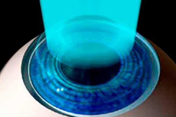
  • Estágio I (inicial) – Óculos e Lentes de Contato Gelatinosa + Crosslinking da córnea
  • Estágio II (inicial a moderado) – Lentes de Contato Rígida + Crosslinking da córnea
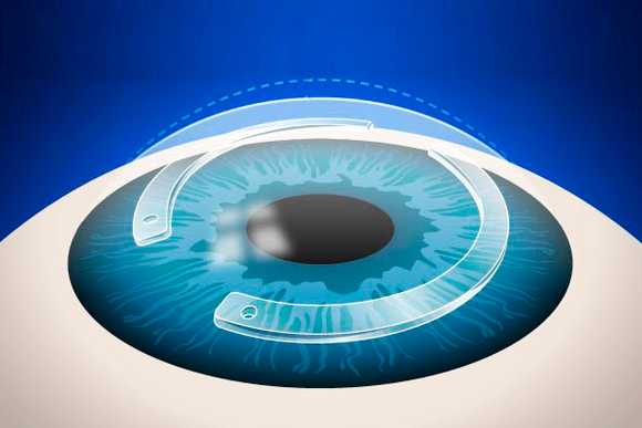
  • Estágio II/III (moderado) – Implante de Anel (indicado somente em casos específicos) + Crosslinking da córnea

Como evitar o Transplante de Córnea?

Imagem Como evitar o Transplante de Córnea?

Assim que diagnosticado o ceratocone, é imprescindível que o paciente faça um acompanhamento de perto com seu médico. Nos estágios iniciais do ceratocone, é fundamental tratar qualquer patologia de pálpebra e conjuntiva associada.

O objetivo é eliminar todo e qualquer microtrauma na córnea, geralmente desencadeado pelo hábito de coçar os olhos.

O uso de colírios lubrificantes e anti-alérgicos ajudam a prevenir a coceira.

Sendo detectada a progressão do ceratocone, mesmo após o controle de todos os fatores externos e locais do olho, é indicado o fortalecimento da córnea com o crosslinking da córnea.

Novidades sobre o Tratamento do Ceratocone

Moderno Centro Especializado no Tratamento do Ceratocone na Av. Paulista, 1499 – 9º Andar